High-Frequency Electron Paramagnetic Resonance
High-frequency EPR in field and frequency domains for investigating paramagnetic and other materials.
Pulsed Q-Band EPR
Pulsed Q-Band EPR to investigate the coherence time and other spin dynamics in molecular quantum bits.
X-Band EPR
CW X-band EPR to characterize the magnetic anisotropy of molecular nanomagnets.
SQUID Magnetometry
SQUID magnetometry to investigate static and dynamic magnetic properties of a magnetic material.
Magnetic Circular Dichroism Spectroscopy
Magnetic circular dichroism spectroscopy to investigate the electronic structure and magnetic properties of a material in one go.
Synthesis
Synthesis to prepare new materials with exciting physical properties.
Ab initio calculations
Ab initio calculations to understand the electronic structure of molecular magnetic materials.
Mößbauer Spectroscopy
Mößbauer spectroscopy to investigate the electronic structure and magnetic properties of iron containing samples, e.g. catalysts of the future.
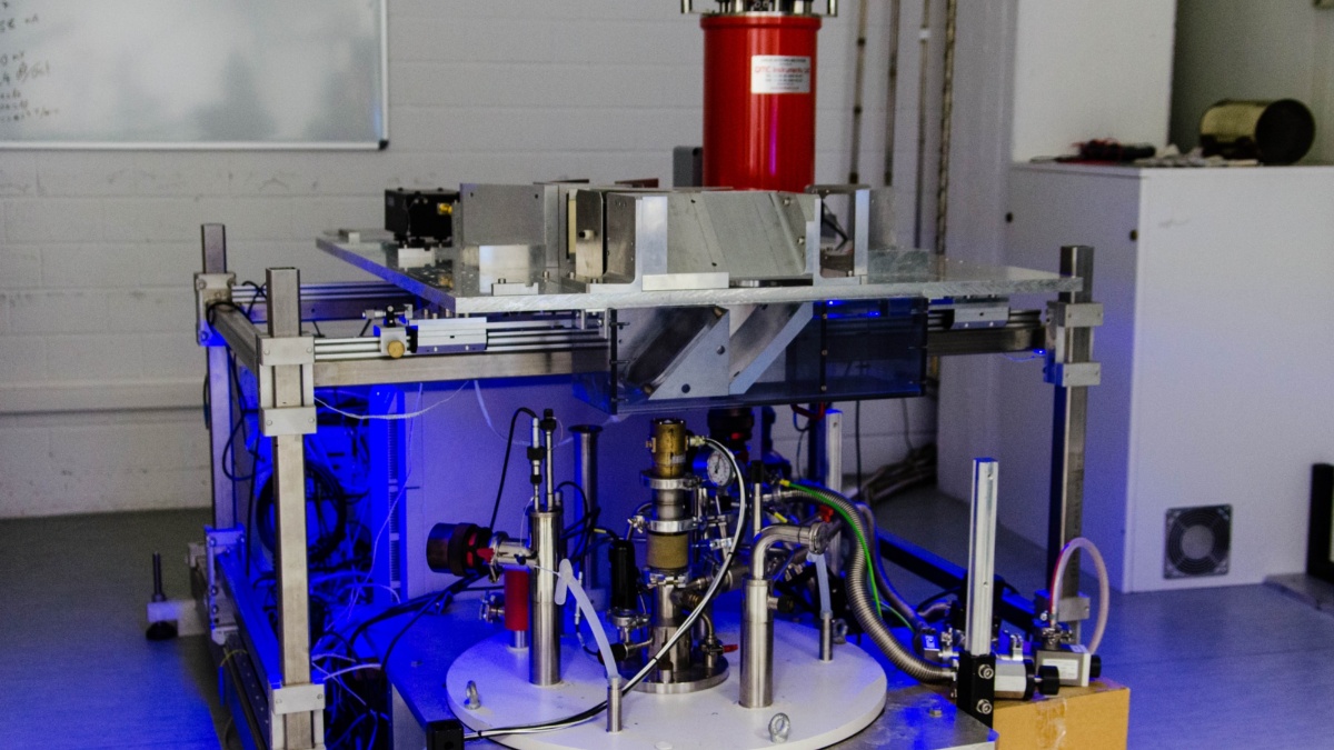
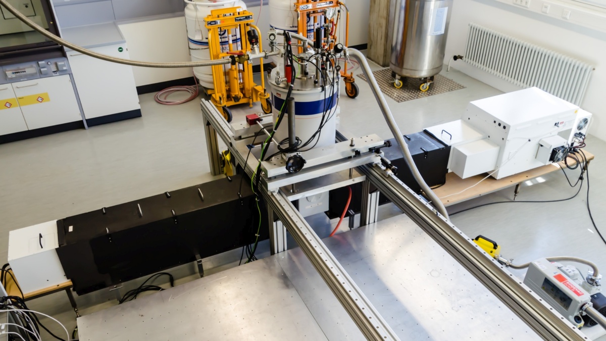
High-frequency Electron Paramagnetic Resonance spectroscopy
We use our home-built broadband HFEPR spectrometer to investigate the magnetic anisotropy of molecular nanomagnets in great detail and elucidate its origins. Our spectrometer operates at frequencies from 85 - 1100 GHz in fields of up to 17 T and temperatures down to 1.5 K. Beyond EPR, we also use this instrument for the investigation of collective magnetic resonance in magnetically ordered systems and for magneto-optical investigations of graphene.
Frequency-domain magnetic resonance
The HFEPR spectrometer can also be operated in the frequency-swept mode, enabling us to measure frequency domain spectra in zero-field and to generate field-frequency excitation maps. By using very fast sweep rates, we can perform rapid scan EPR measurements to gain access to spin dynamics at very high frequencies. For higher excitation energies we use a FTIR spectrometer in the THz-FIR frequency domain, especially for highly anisotropic systems including lanthanide complexes.
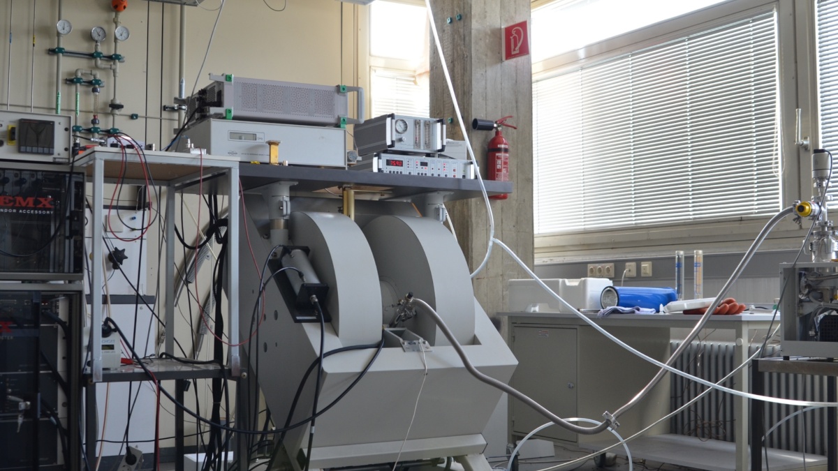
CW X-Band EPR, EDMR
EPR at 9.5 GHz (X-band) enables us to investigate and precisely determine weak interactions in molecular nanomagnets. By recording the influence of magnetic field and microwave irradiation on the electrical transport of a semiconducting sample, we can learn about the properties of mobile charge carriers (electrically detected EPR).
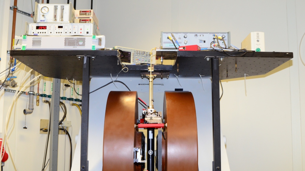
Pulsed Q-Band EPR
We use our pulsed Q-Band (35 GHz) EPR spectrometer to investigate the spin dynamics. We are especially interested in the quantum coherence of molecular quantum bits and the factors influencing it. The spectrometer can also perform electron-nuclear double resonance (ENDOR) measurements to investigate the coupling between electron and nuclear spins.
MCD and Luminescence
Magnetic circular dichroism is the difference in absorption between left- and right-hand circularly polarized radiation and yields information on both the magnetic properties and electronic structure of molecular nanomagnets. Our homebuilt spectrometer operates from 5000 to 40000 cm-1 in fields up to 10 T and at temperatures down to 1.5 K. We employ luminescence spectroscopy in the UV/Vis/NIR range as a complementary technique to study the electronic structure of molecular magnetic materials.
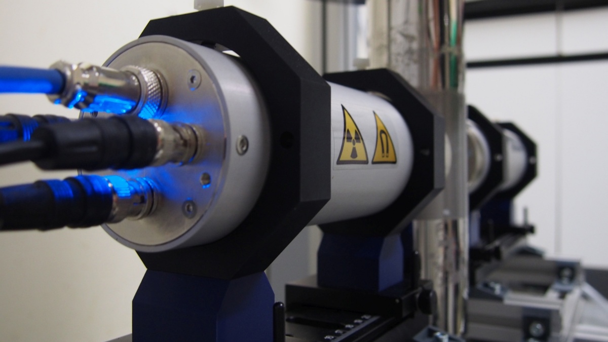
Mößbauer spectroscopy
Mößbauer spectroscopy is a very sensitive probe to study oxidation state and electronic structure and dynamics of iron-containing samples. Our spectrometer is a variable-temperature setup that operates down to 4K.
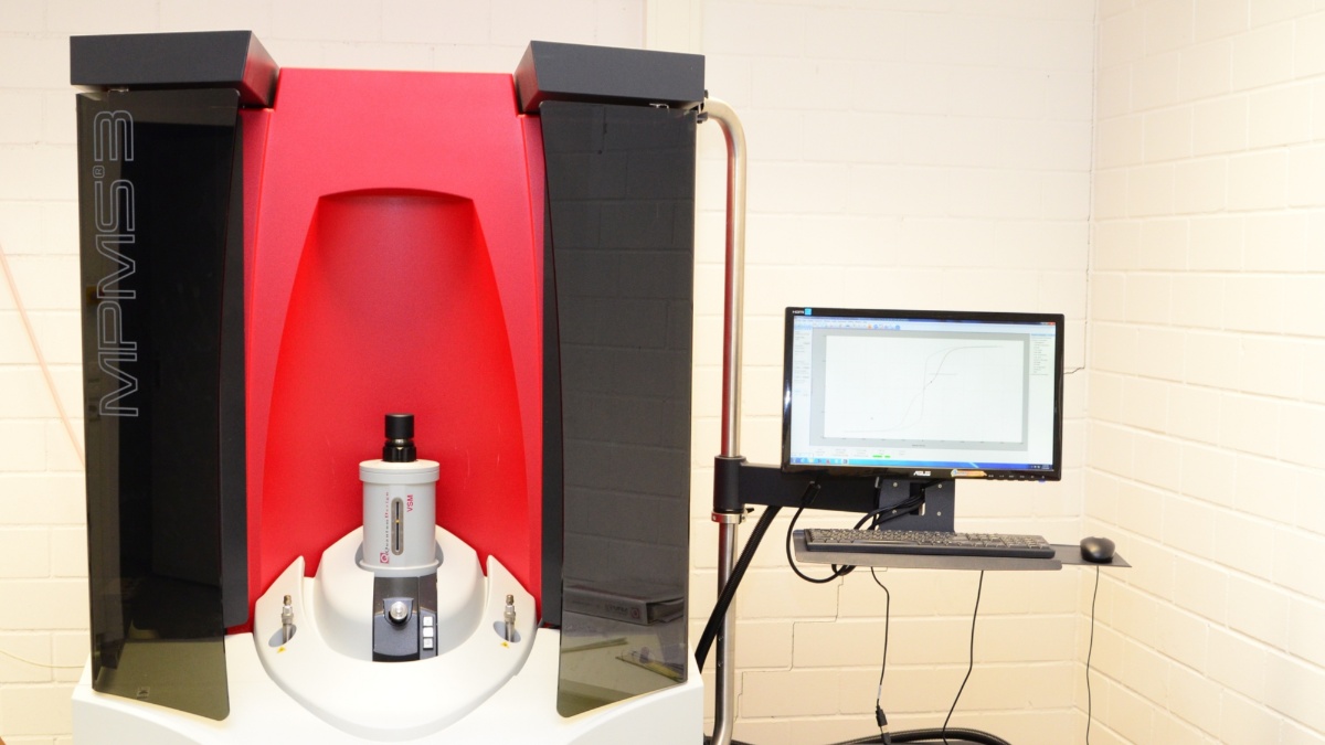
SQUID magnetometer
Magnetometry is a standard method to investigate the spin centers, their dynamics and their interactions. Jointly with Inorganic Chemistry department, we operate a Quantum Design MPMS3 SQUID magnetometer with AC, transport and light irradiation capabilities.
AFM
Atomic force microscopy allows us to structurally characterize 2D materials such as graphene as well as thin layer samples.
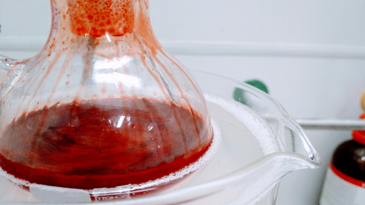
Synthesis
We have a spacious synthesis laboratory with four fumehoods, vacuum lines and a glove box. This allows us to synthesize novel materials for our studies, such as lanthanide single molecule magnets and molecular 2-qubit systems.
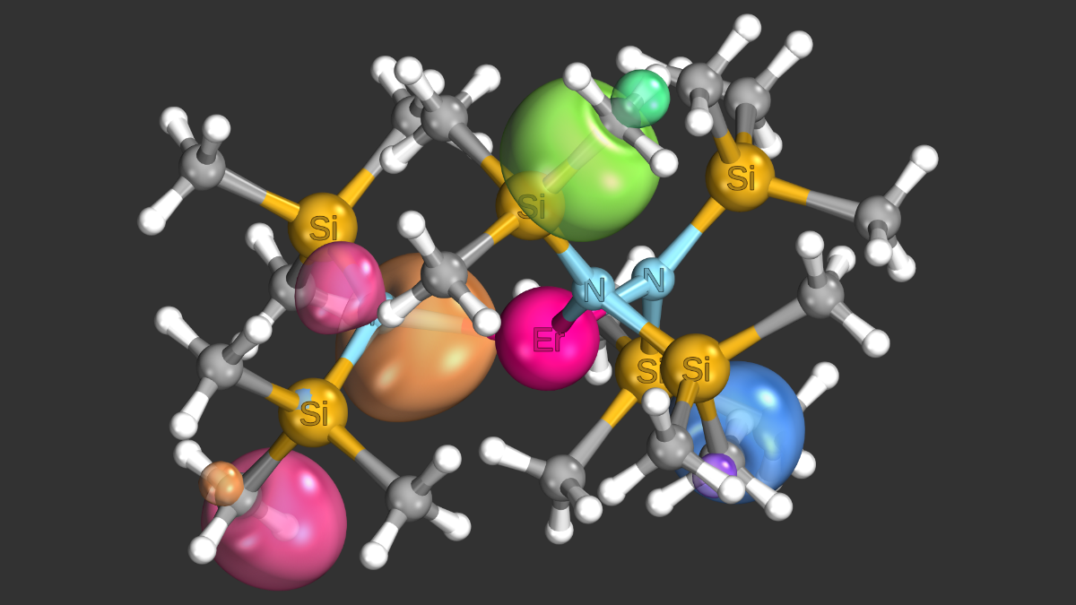
Ab initio calculations
Multireference ab initio calculations are essential to understand the electronic structure of molecular magnets. We have developed new methods to improve speed and accuracy of such calculations, the latter involving better descriptions of dynamic electron correlations.