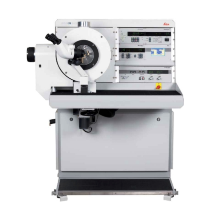
The Freeze Fracture and Etching System BAF060 [1] is used for the preparation of different specimens from various samples the structure of which are to be investigated. Emulsions, suspensions, liquids, gels, biological samples, etc. can be prepared with this instrument to visualize their structure. Freeze-fracture replication and freeze etching techniques involve the production of ultrathin heavy metal films (replicas) from a fractured surface of a frozen specimen. Freeze etching provides more information from the fracture faces by subliming superficial ice layers under vacuum to expose some elements that were originally hidden. Generally, the replica is examined in a conventional transmission electron microscope. Another method is to study the topography of the coated sectioned surface in a scanning electron microscope combined with cryo-stage.
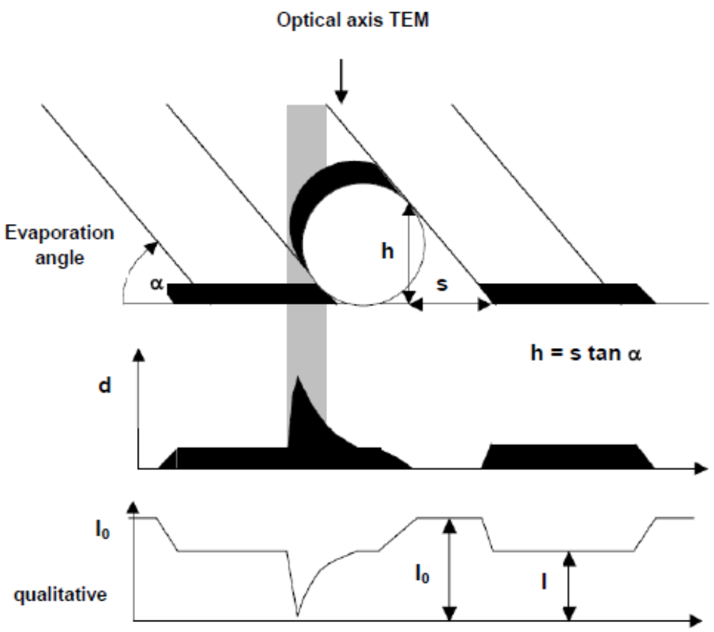
Before using the BAF060 the sample has to be frozen on a special specimen carrier (for example by plunge-freezing, cryo-jet, high pressure freezing, etc.). The fracturing of frozen specimens is then carried out in a precooled vacuum chamber with a microtome (LN2 cooled fracturing knife with manual or motorized motion controlled with stepper motor) or by using fracturing device for sandwiches (double-replica specimen table). Different options for the coating process with Pt/C and C are possible. The electron beam source angles can be preset from 0 to 90° for shadowing and replication (carbon layer at 90° to stabilize the metal film). Furthermore, stationary or rotary shadowing is possible, offering a wide range of shadowing options, including DARS (Double Axis Rotary Shadowing). The film thickness is precisely controlled with automated quartz crystal thickness monitor and shutter termination. As shown in Fig. 1-2, shadowing is a way to reveal the shape of the fracture or of particles because it introduces some mass contrast to the thickness contrast image. After freeze fracture and shadowing has been completed, the heavy metal/carbon replicas must be thawed out and cleaned from adhering chemical/biological material before examining with TEM.

|
[2] Freezing, Freeze-Etching and Replica-Production for Electron Microscopy, U. Zeile, BAL-TEC EM Technology and Application, 2000. |
|
[3] Transmission Electron Microscopy, D. B. Williams, C. B. Carter, Springer, Amsterdam, 2009. |

Natalie Preisig
Dr.Scientific Staff